Fluorescence Microscopy With Multiple Fluorochromes. Fluorescence imaging in its conventional form reveals biological structures and processes but is limited by the number of fluorochromes that can be distinguished in one measurement. Fluorescence With Multiple Fluorochromes This interactive tutorial explores the effects of fluorescence cube filtration of excitationemission spectra on specimens stained with multiple fluorochromes. Current filter sets from Carl Zeiss 2. Filter set in modern filter modules Contents with light trap 1.

By combining antibodies that are labeled with different fluorochromes the location of several proteins can simultaneously be visualized in one sample. Fluorescence With Multiple Fluorochromes This interactive tutorial explores the effects of fluorescence cube filtration of excitationemission spectra on specimens stained with multiple fluorochromes. Current filter sets from Carl Zeiss 2. A computer generates the image from this fluorescence data. Immunofluorescence microscopy is a unique method to reveal the spatial location of proteins in tissues and cells. Although the fluores-cence microscope cannot provide spatial resolution below the diffraction limit of the respective objects the de-.
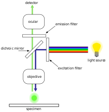
Around 1962 Ploem started work in collaboration with Schott on the development of dichroic beamspitters for reflection of blue and green light for fluorescence microscopy using epi-illumination.
These flourchromes when exited with fluorescent light of given wavelength emit light with longer wavelength. Fluorescence Microscopy Fluorescence microscope uses fluorescent light to excite fluorescent specimens. Despite the numerous advances made in fluorescent dye synthesis during the past few decades there is very little solid evidence about molecular design rules for developing new fluorochromes particularly with regard to matching absorption spectra to available confocal laser excitation wavelengths. These flourchromes when exited with fluorescent light of given wavelength emit light with longer wavelength. Around 1962 Ploem started work in collaboration with Schott on the development of dichroic beamspitters for reflection of blue and green light for fluorescence microscopy using epi-illumination. Fluorescence microscopy reveals the distribution of fluorochromes in biological specimens whether they are introduced externally as probes or occur naturally as chlorophylls and porphyrins.