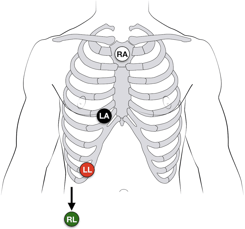
Bedside 3 Lead Ecg Placement. These colors are not universal as two coloring standards exist for the ECG discussed below. These are the most common 3 lead ECG placements. Placement of Lead V4 V4 should be placed before lead V3. The limb lead is shown in red negative and yellow positive electrodes.

3 lead ECG cable Placement there are two ways Way 1. Discuss the troubleshooting procedures for inaccurate heart rate and arrhythmia detection and. Each electrode corresponds with a single ECG lead unlike the limb electrodes. 3 and 5-lead monitoring take electrodes that are used for the limbs in 12-lead monitoring and instead place them on the chest wall in order to reduce artifacts ECG signals that are from sources other than the heart caused by patient movement Khan 2015. Patient factors may also contribute to the variability in accuracy such as the patients respiration position smoking recent dietary intake and obesity McCann et al. Discuss procedures for ECG electrode placement and skin preparation.
Just so where do you put ECG leads.
Discuss the troubleshooting procedures for inaccurate heart rate and arrhythmia detection and. Deviation of lead placement even by 20-25mm from the correct position can create clinically significant changes on the ECG including changes to the ST-segment McCann et al. Placed the red electrode within the frame of rib cageright under the clavicle near shoulder see chart in follow picture LA. Right arm limb lead is white white goes to the right forearm proximal to the wrist. The yellow electrode is placed below left clavicle which is in the same level of the Red electrode. 3 lead ECG cable Placement there are two ways Way 1.